Sensational How To Read Obstetric Sonography Report

Basically the educational path to become an obstetric sonographer starts with training in the field of diagnostic medical sonography.
How to read obstetric sonography report. The reflected sound waves are then picked up by the ultrasound transducer. Fetal biometric parameters are antenatal ultrasound measurements that are used to indirectly assess the growth and well being of the fetus. Based on menstrual dataearly scan.
It does not use ionizing radiation has no known harmful effects and is the preferred method for monitoring pregnant women and their unborn babies. The procedure is a standard part of prenatal care in many countries as it can provide a variety of information about the health of the mother the timing. Its most important use however is to determine anomalies andgenetic disorders in the foetus early on in pregnancy that gives time for the doctors and the expectant mother to think about corrective measures even while the baby is in the womb.
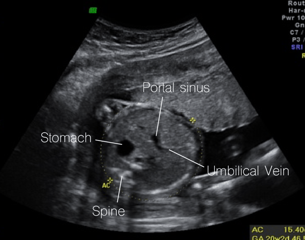
Of ultrasound examination as it may contain additional documen-tation requirements. AIUM Practice Guideline for the Performance of Obstetric Ultrasound Examinations 2007 o Irregular menstrual periods ¾ If the mother has had irregular menstrual periods in the year prior to the current pregnancy then one ultrasound can be performed to confirm dates report one. Fetal ultrasound measurements can include the crown-rump length CRL biparietal diameter BPD femur length FL head circumference HC occipitofrontal diameter OFD abdominal.
These sound waves are then reflected by different tissue types in different ways. 3cm Vertical Fluid Pocket. The sound waves are then transformed into an image by special software.
Because sonography is not 100 sensitive Color Doppler ultrasound findings may be normal blood flow maintained despite the presence of ovarian torsion. Requirements for the Ultrasound Examination Ultrasound examinations should be recorded in a manner that will allow subsequent review for adequacy for diagnostic purposes. Qualities of an Obstetric Sonographer.
Ultrasound EDC if gestation age is 21 weeks. For external reports the facility name and contact details should be clearly stated. There is a focal anechoic tear of the anterior distal aspect of the supraspinatus tendon measuring 1 cm short axis by 15 cm long axis.


